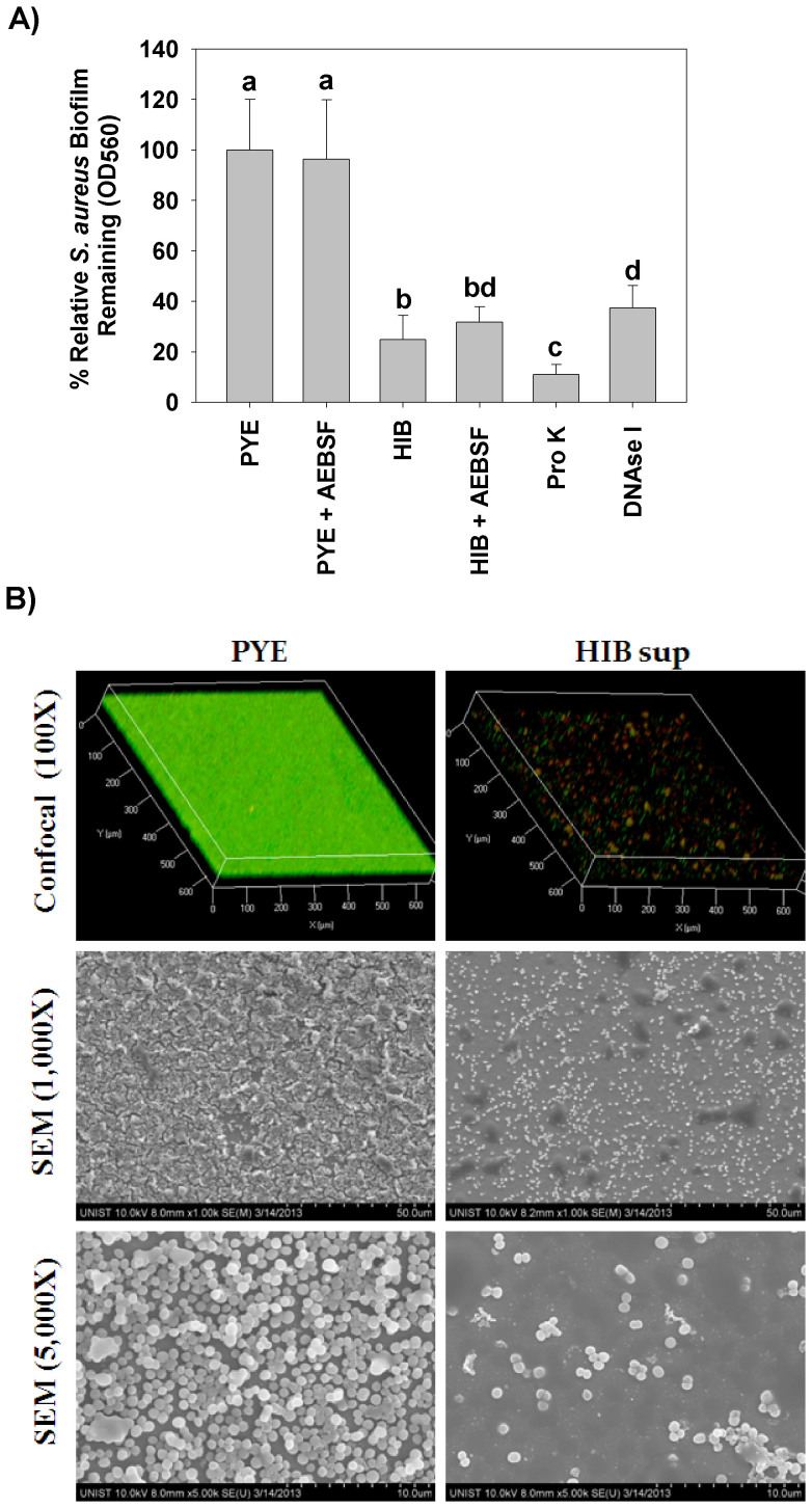
Figure 3.
(A) Removal of S. aureus biofilms by HIB supernatants, proteinase K and DNAse I. S. aureus biofilms were prepared in 96 well plates. Afterwards, the medium was removed, and the wells were filled with 200 μl of HEPES buffer supplemented with either 10% PYE medium, 10% HIB supernatant, 100 μg/ml proteinase K or 20 μg/ml DNAse I. The plates were then incubated for 24 h more, washed and CV stained (a, b, c, and d = P < 0.05). (B) Confocal and SEM images for the S. aureus biofilms on silicon chips with and without HIB supernatant treatment. S. aureus biofilms were formed on silicon chips, washed and incubated for 24 h with HEPES buffer supplemented with either 10% PYE medium or HIB supernatant. The chips were then washed and analyzed. For the confocal microscopy(Upper photos), the biofilms were stained with CFSE (green) for the live cells and EthD-1 (red), for the dead cells and extracllular DNA. The middle and the lower photos show the SEM images for the chips at low and high magnifications, respectively.

