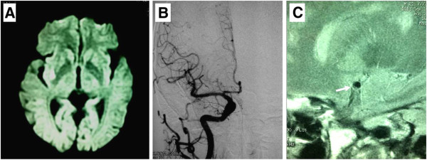
Figure 1.

MRI, DSA and HR MRI findings of Case 1. (A) MRI diffusion weighted image shows a hypersensitive lesion on right putamen with rostral extension to the corona radiates. (B) Digital angiography of the right MCA shows normal. (C) High-resolution MRI of the MCA demonstrates an atherosclerotic plaque (arrow) on the frontal wall of the right proximal MCA.
