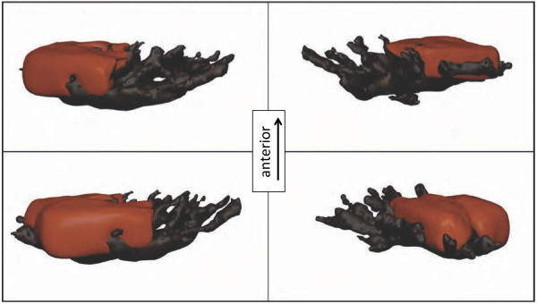
Figure 8.

3D-Model of a reconstructed fluorescent chromatophore unit with one melanophore (black) embracing four fluorescent globular iridophores (red, two shown, two others omitted). Sample taken at an intermediate state with not yet fully aggregated melanosomes.
