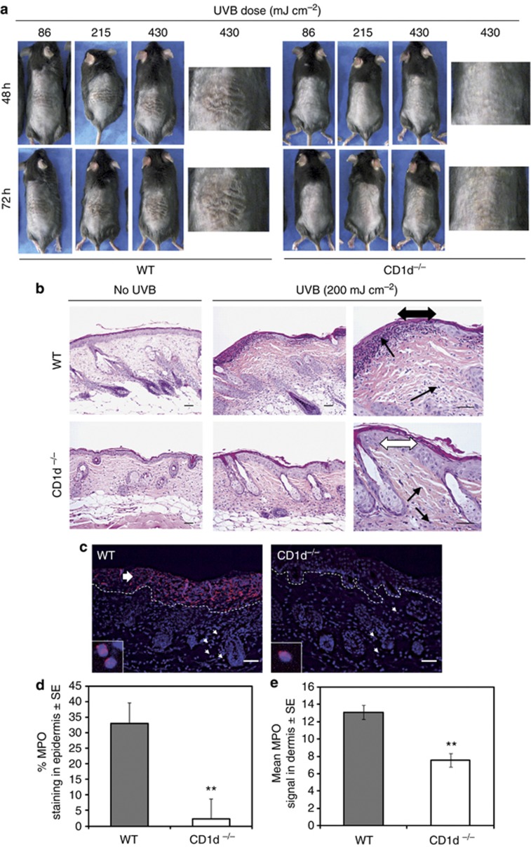
Figure 1.
UVB-induced cutaneous tissue injury and inflammation are abolished in CD1d-knockout mice. (a) Photographs of single UVB-irradiated backs of CD1d knockout (CD1d−/−) and wild-type (WT) (C57BL/6 × 129) mice exposed to different UVB doses. (b) Hematoxylin and eosin staining of the backs of single UVB-irradiated C57BL/6 CD1d−/− and WT mice at 48 hours. Single arrowheads: inflammatory infiltrates; double arrowheads, black: epidermis erosion, white: intact epidermis. (c) Immunohistofluorescence of skin sections from (b) stained for myeloperoxidase (MPO) and 4,6-diamidino-2-phenylindole (DAPI). Dashed: epidermis–dermis junction. Small arrows: MPO-positive cells. Large arrow: epidermis ulceration. (d) Mean epidermal MPO staining (normalized to the total epidermal surface)±SE for WT (n=3) and CD1d−/− (n=4) skin sections; analysis of variance, **P<0.01. (e, d) Mean MPO-positive cells in the dermis (per 104 μm2)±SE. Scale bar=0.05 mm.

