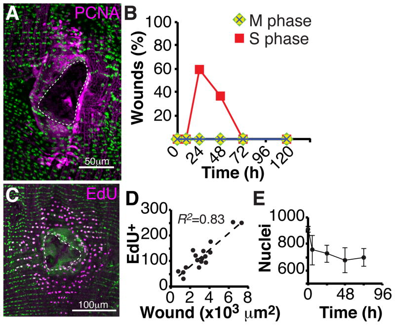
Figure 2. Epithelial cells re-enter the cell cycle, but do not divide.
(A) Immunofluorescent image of PCNA-GFP (magenta) expression at 24 hours post injury. DAPI (green); Scar (dashed white line). (B) S phase, but not M phase cell cycle markers are expressed post injury. Time course of the percentage of wound zones with cells expressing the indicated cell cycle markers. S phase marker (PCNA-GFP, red square) and M phase markers (PH3, green diamond; CycB-GFP, yellow circle; Polo kinase-GFP, blue cross). N=30 wounds/ time point. (C) Immunofluorescent image of EdU-positive nuclei (magenta) within the wound zone following continuous EdU labeling and analysis at 3 days post injury. Epithelial nuclei (green, flpout nlsGFP; Epithelial-Gal4/ UAS-Flp); Scar (dashed white line). (D) The number EdU-positive nuclei correlates with wound area (N=17). (E) Nuclei number is not restored following epithelial repair. Time course of the abdominal epithelial nuclear number within a fixed zone (7.5 × 104 μm2) following injury at the zone’s center (N=5, mean ± SD).

