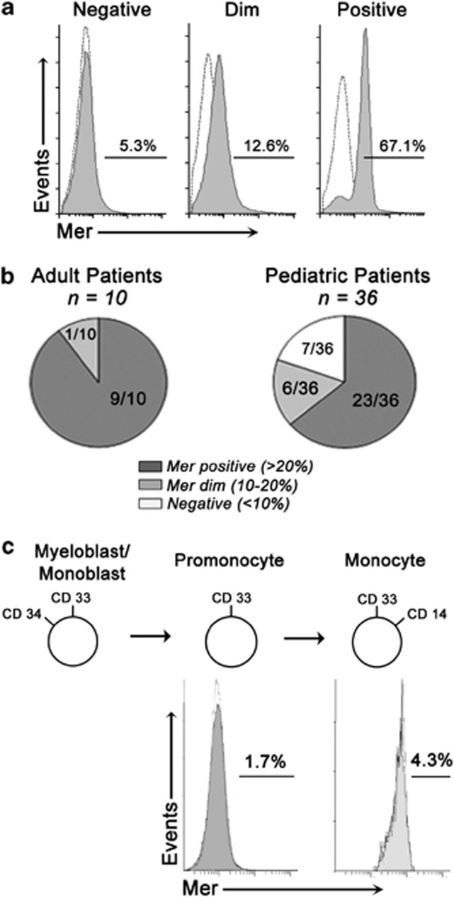
Figure 2.
Mer is expressed in a majority of diagnostic bone marrow samples derived from patients with AML, but not in normal bone marrow. Myeloblasts collected at diagnosis from patients with AML were analyzed by flow cytometry for Mer expression. (a) Cells that expressed human CD33 and CD45 were analyzed after staining with a mouse anti-human Mer antibody and PE-conjugated secondary antibody to determine Mer expression (gray histogram). Non-specific staining was determined using a mouse IgG1 isotype control antibody and PE-conjugated secondary antibody (white histogram). Representative flow cytometry profiles for Mer-positive (>20% cells positive), Mer-dim (10–20% cells positive) and Mer-negative (<10% cells positive) patient samples are shown. (b) Graphic representation showing the fractions of Mer-positive, Mer-dim and Mer-negative pediatric (right panel) and adult (left panel) diagnostic primary patient samples. (c) Bone marrow samples from eight healthy donors were evaluated by flow cytometry for Mer RTK expression as described above. Myeloid progenitor populations were identified by gating on the cell surface markers noted in the figure. The fractions of cells expressing Mer in progenitor populations are shown. Myelo/monoblastic populations could not be quantified, due to low-cell number.

