Figure 1.
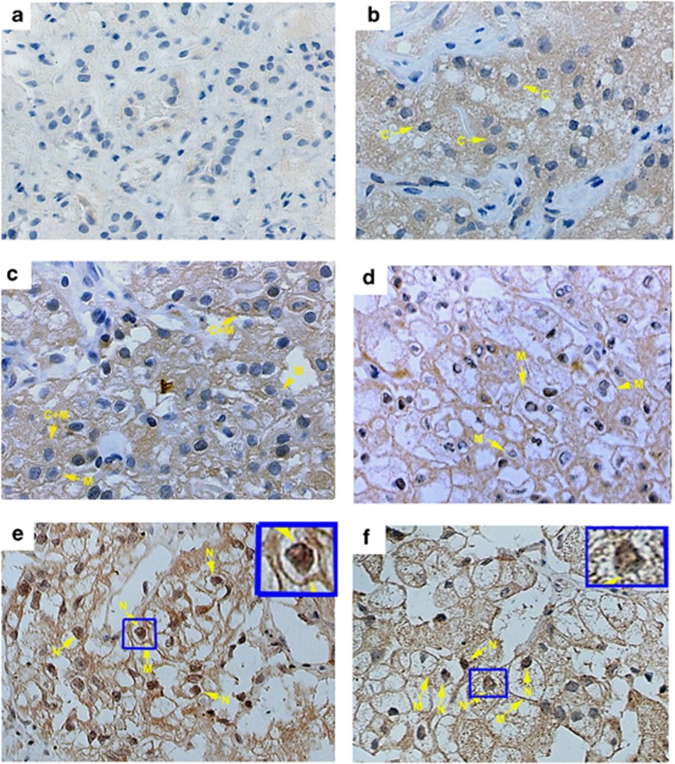
Representative immunohistochemical staining of B7-H4 in RCC tissues using anti-B7-H4 mAb 3C8. (magnification × 400) (a) Negative control. (b) B7-H4 was predominantly expressed in the cytosol. (c) B7-H4 was predominantly expressed on the membrane and in the cytosol. (d) B7-H4 was predominantly localized on the membrane. (e) and (f) B7-H4 was predominantly localized on the membrane and in the nucleus.

