Fig. 1.

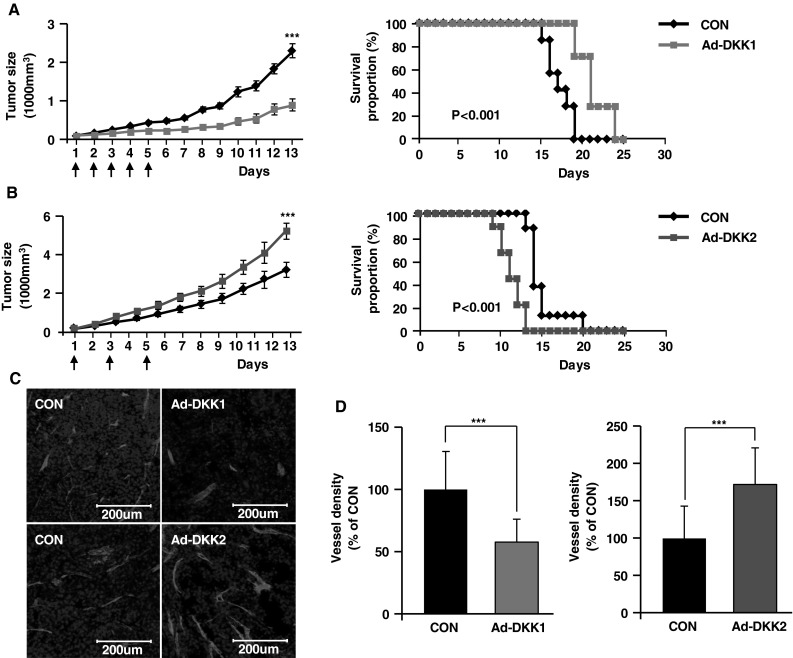
Adenoviral DKK1 and DKK2 expression differentially modulate B16F10 melanoma growth and angiogenesis. B16F10 murine melanoma cells were injected subcutaneously into abdomens of C57BL/6 mice. Mice were injected intratumorally with control adenovirus (CON, n = 8), DKK1-expressing adenovirus (Ad-DKK1, n = 8), or DKK2-expressing adenovirus (Ad-DKK2, n = 8) on the indicated days (left panels, vertical arrows). Tumor growth was monitored over time, and data are presented as mean ± SE. The percentage of surviving mice was determined by monitoring the tumor growth-related events (tumor size >3,000 mm3) over a period of 25 days (a, b, right panels). 1,000 mm3 adenovirus-infected tumors were stained with anti-CD31 antibody, an EC marker. By confocal imaging of tumor sections, vessel density was determined (c) and quantified (d). Nuclei were stained with DAPI. Data are mean ± SD (***p < 0.001)
