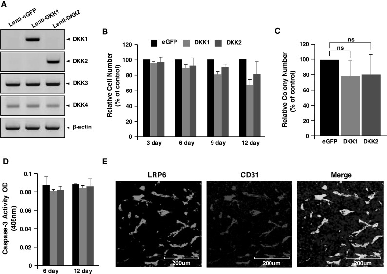
Fig. 3.

DKK1 or DKK2 expression does not alter tumor cell proliferation, colony formation, or apoptosis. a B16F10 tumors were stably transfected with GFP-, DKK1-, or DKK2-expressing lentivirus. Protein levels of DKKs (1–4) were detected in transfectant lysates by western blotting. β-actin was used as a protein loading control. b Cell numbers were determined in relation to GFP-expressing cells (set at 100 %) at the indicated times. c Colony-forming efficiency of the stable transfectants at 3 weeks after seeding. Data show the number of soft agar colonies relative to GFP-expressing cells (set at 100 %). d Stable transfectants were cultured for 6 or 12 days, and caspase-3 activity was measured as an indicator of apoptosis. e B16F10 melanomas generated in wild-type mice were frozen-sectioned and immunostained with anti-LRP6 and anti-CD31 antibodies. Data are mean ± SD of triplicate experiments (ns not significant; p > 0.05)
