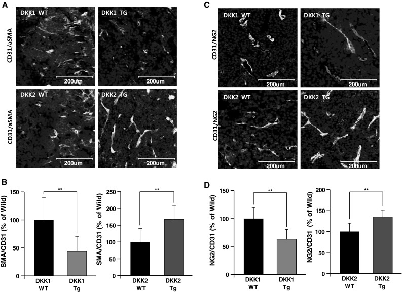
Fig. 6.

Perivascular cell coverage of tumor vessels is inhibited by DKK1 and enhanced by DKK2. Sections of B16F10 melanoma tumors generated in DKK1 Tg, DKK2 Tg, and wild-type mice were stained with anti-CD31 and anti-smooth muscle actin (SMA) antibodies. Nuclei were stained with DAPI. SMC coverage is presented (a) and quantified as the ratio of SMA+ area to CD31+area (b). Sections of tumors generated in DKK1 Tg, DKK2 Tg, and wild-type mice were stained with anti-CD31 and anti-NG2 (pericyte-specific) antibodies. Pericyte coverage is presented (c) and quantified as the ratio of NG2+ area to CD31+ area (d). Nuclei were stained with DAPI. Data are mean ± SD (*p < 0.05; **p < 0.01)
