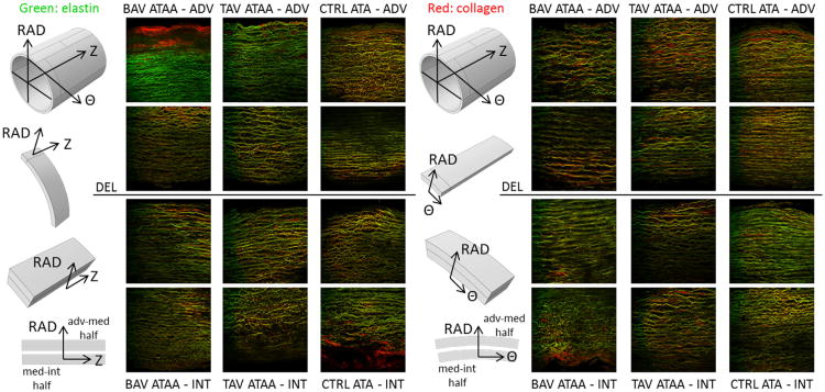
Fig. 1.
Example full wall thickness multi-photon microscopy images (0. 5 × 0.5 mm2 each) of elastin (green) and collagen (red) fibers in the Z-RAD (columns 2-4) and Θ-RAD planes (columns 6-8) of BAV-ATAA (columns 2,6), TAV-ATAA (columns 3,7) and CTRL-ATA (columns 4,8). Column 1: The adventitial-medial and medial-intimal delaminated halves from Θ strips were placed under the microscope with the Z-RAD plane facing up. Column 4: The adventitial-medial and medial-intimal delaminated halves from Z strips were placed under the microscope with the Θ-RAD plane facing up. Rows 1,2: The adventitial-medial delaminated half. Rows 3,4: The medial-intimal delaminated half.

