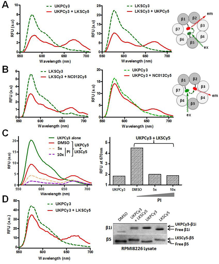
Fig. 5.
FRET signals detected from the purified constitute proteasome (A–C) treated with FRET pairs: A. UKPCy3:LKSCy5 (left) and LKSCy3:UKPCy5 (right); B. LKSCy3:NC012Cy5 (left) and UKPCy3:NC012Cy5 (right). C. FRET signals were attenuated by co-treatment with an excess of non-fluorescent proteasome inhibitors (PI). RFU: Relative fluorescence units. D. FRET signals detected from RPMI multiple myeloma cell lysates treated with UKPCy3:LKSCy5 pair (Left). When UKPCy3 was co-incubated with LKSCy5 in RPMI8226 cell lysates, UKPCy3 selectively labeled β1i, whereas LKSCy5 selectively labeled β5.

