Figure 5.
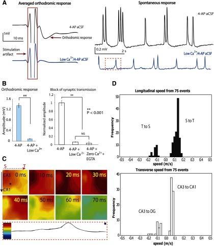
Propagation is independent of synaptic transmission. A, Left, Inhibition of synaptic transmission by low Ca2+/4-AP solution perfusion. Orthodromic responses were recorded from pyramidal cells in CA3. The averaged responses shown here are 25 responses from 7 different experiments, for both 4-AP and low Ca2+/4-AP condition. Right, The change of the amplitude of spontaneous responses before and after applying low Ca2+/4-AP solution. B, Left, Synaptic transmission is blocked by low Ca2+/4-AP aCSF. Orthodromic response amplitudes decreased significantly with the perfusion of low calcium solution (25 samples). Data are mean ± SE. **p < 0.001. NS, Not significant. Right, A similar suppression of orthodromic response was observed with perfusion of zero calcium/4-AP aCSF and EGTA (50 μm). C, Wave propagation maps from all 64 channels in the same time window (marked in dashed box in A). Bottom, Coding to the color bar. In this example, the septal side is on the left and temporal side is on the right. Spontaneous activity in low Ca2+/4-AP aCSF initiated in the septal area of CA3 and propagated longitudinally. From 40 ms onward, the activity propagated longitudinally to the temporal side. D, Histograms of the speed recorded in all four directions from 7 different experiments.

