
Figure 7.
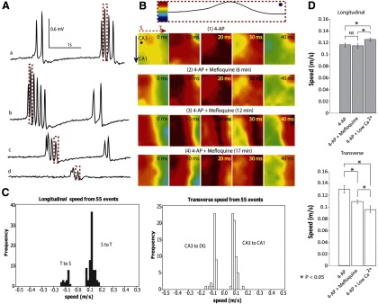
Propagation does not require gap junctions. A, Effect of mefloquine on the amplitude of 4-AP-induced spontaneous activity. Mefloquine has been shown to block the conduction of gap junctions effectively within 3 min (Cruikshank et al., 2004). Signals were obtained from one channel (the dot shown in B) in the array at different times as the tissue was incubated in mefloquine aCSF with 4-AP for >3 min. a, 4-AP aCSF only; b, after perfusion of mefloquine/4-AP aCSF for 6 min; c, after applying mefloquine/4-AP aCSF for 12 min; d, after applying mefloquine/4-AP aCSF for 17 min. The amplitude of the activity decreased with time. B, Top, The normalization of the peak from dashed box in A. The four rows of color maps (1–4) correspond to the peaks highlighted in the dashed boxes in A. Although the amplitude decreased, blockade of gap junctions did not stop the propagation of activity. C, Propagation speed histogram of the spontaneous activity from two unfolded hippocampus preparations after perfusion with mefloquine. The activity still traveled from the septal to temporal side but also traveled from temporal to septal side in a minority of cases. In the transverse direction, the source was always in CA3 and could travel from CA3 to CA1 or CA3 to DG depending on the location of the source. D, 4-AP-induced epileptiform activity was analyzed by separating transverse versus longitudinal propagation directions. Plots show the speed (mean ± SE) in both longitudinal (within CA3) and transverse directions. In the longitudinal direction (top), low Ca2+/4-AP aCSF did speed up propagation, whereas in the transverse direction (bottom), both mefloquine and low-Ca2+ solutions decreased the speed. *p < 0.05.