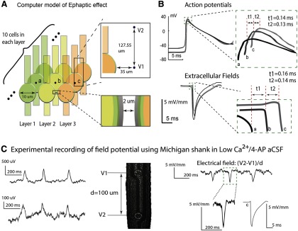
Figure 8.

Propagation speed is consistent with electrical field-mediated transmission. A, A computer model was constructed to simulate a Ca2+-free, electrical field communication-only environment. The extracellular voltages V1 and V2 were measured at two points outside of the middle cell in each row. Top inset, The measurement for the center cell c in layer 3. Extracellular field is calculated by finding the spatial first difference of these two extracellular voltages. B, Top, Membrane potentials of the middle cell in each row. All cells in the first row were stimulated by an experimentally acquired extracellular signal. We observed a propagation speed of 0.086 m/s between the center cell (a) of the first row and the second row, and 0.092 m/s between the center cell (b) of the second row and that (c) of the third row. Bottom, Extracellular field waveforms outside of the center cells (a–c) in each row. The amplitudes of the three fields are 7.05, 5.77, and 5.05 mV/mm. The time delays between the first and second and between the second and the third row are 0.16 ms and 0.14 ms, respectively, and the corresponding propagation speeds are 0.075 m/s and 0.086 m/s. C, Experimental recording of field potential from Michigan electrode (left) and relative extracellular electrical field (right) in an unfolded hippocampus. The field amplitude ranges from 3 to 6 mV/mm, and the values are similar to those obtained computationally and shown in B. These results indicate that an electrical field effect could be responsible for propagation with an approximate speed of 0.1 m/s.
