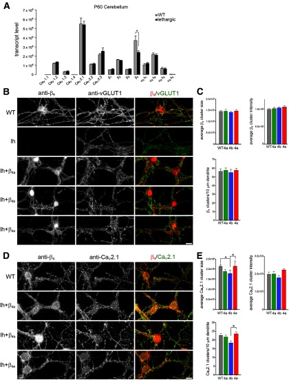
Figure 6.
Expression and distribution of β4 subunits in lethargic cerebellar granule cells reconstituted with β4a, β4b, or β4e. A, Quantitative RT-PCR revealed similar expression of CaV subunit isoforms in WT and lethargic adult cerebellum (mean ± SEM, n = 3). B–E, Cultured cerebellar granule cells from lethargic mice were reconstituted by lentiviral transfection with one of the β4 splice variants, βA-β4a, βA-β4b, or βA-β4e, and immunolabeled with anti-β4, anti-vGLUT1, or anti-β4 and anti-CaV2.1 at DIV 9. B, Wild-type cultures of cerebellar granule cells express β4 subunits in discrete clusters on the processes and around the somata; no expression of β4 subunit was detected in cultures from lethargic mice. C, Quantitative analysis of reconstituted lethargic cultures shows that staining of β4a, β4b, and β4e in the processes is similar to β4 staining in WT controls. However, the three β4 splice variants differ in their localization in nuclei. D, All three β4 splice variants show a similar overall distribution pattern and partial overlap with synaptic CaV2.1 clusters in the soma and along the processes (representative images of 3–4 cultures). E, CaV2.1 cluster size, average cluster intensity, and cluster density along the dendrite were similar to wild-type and to each other, except that size and density of CaV2.1 clusters were reduced upon β4b reconstitution. *p < 0.05, ANOVA and Tukey post hoc analysis. Scale bar, 10 μm.

