Figure 3.
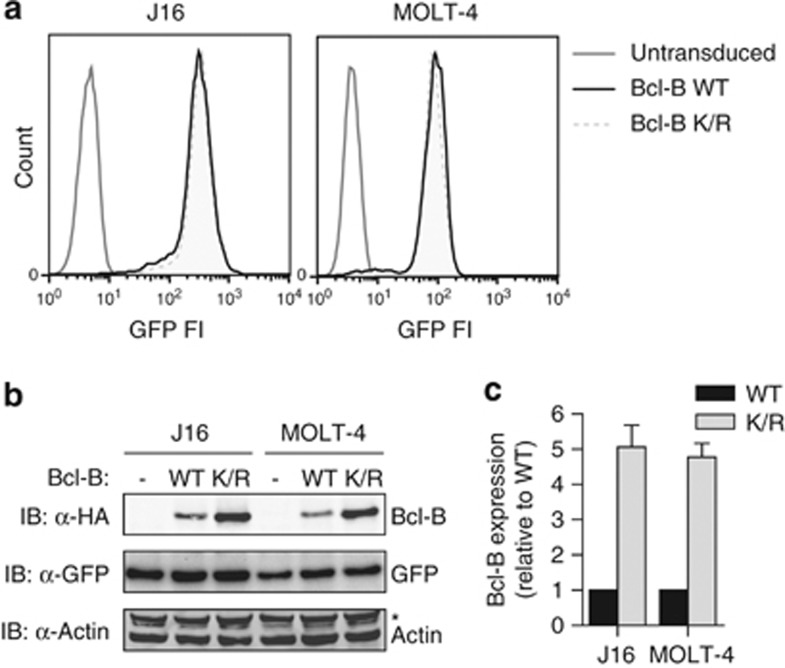
Steady-state protein expression of Bcl-B is regulated by ubiquitination. (a) MOLT-4 and J16 cells were retrovirally transduced with an internal ribosomal entry site (IRES)–GFP vector to express N-terminally HA-tagged Bcl-B in WT or lysineless K/R mutant form. In addition, control cells (- in panel b) were created by transduction with empty IRES–GFP vector. Cells were flow cytometrically sorted twice for equal GFP expression. Histograms depict GFP fluorescence intensity of the different cell lines after the second sort. (b) Total cell lysates of the cell lines described in a were analyzed by immunoblotting for protein expression of Bcl-B (anti-α-HA), GFP and actin. The asterisk indicates a background band of unknown nature. Data are representative of three independent experiments. (c) Quantification of Bcl-B protein expression levels, as determined in b. Bcl-B signal intensity was corrected for GFP signal intensity in the same cells and Bcl-B WT expression level was set to 1. Data represent mean +s.d. from three independent experiments.

