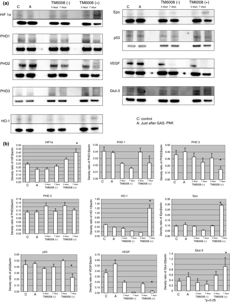
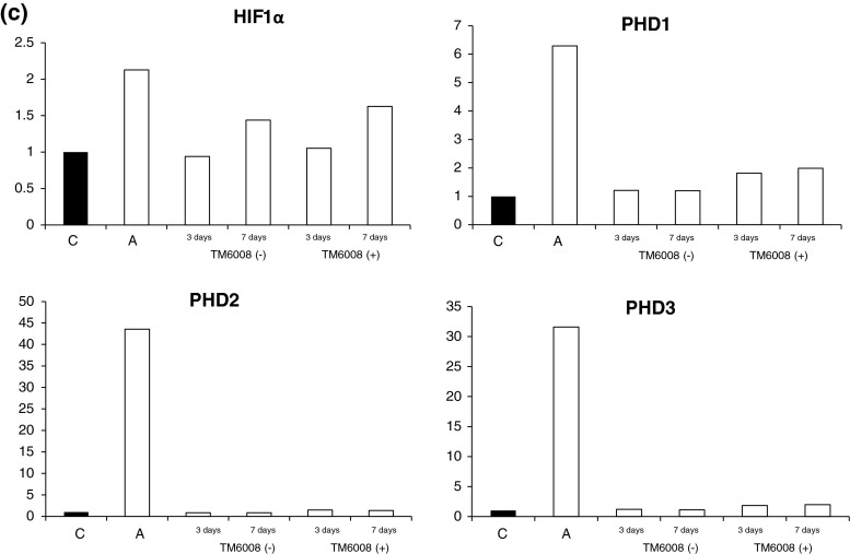
Fig. 3.

Immunoblottings for HIF and molecules downstream of HIF. (a) The protein expression of HIF in cells cultured in the presence of TM6008 at seven days after hypoxia was augmented, compared with that in cells cultured under other conditions. On the other hand, the protein expression of PHD1 in cells cultured in the presence of TM6008 at seven days after hypoxia was decreased, compared with that in cells cultured under other conditions. The protein expressions of PHD2 and PHD3 were increased, similar to the results for HIF. (b) The protein expression level of HIF was significantly augmented after 7 days of culture in the presence of TM6008, compared with that in the control. The expressions of HO-1 and Epo were similar to the results for HIF. On the other hand, the protein expression levels of PHD2 and p53 were significantly lower after 7 days of culture in the presence of TM6008. 1 Control, 2 just after hypoxia, 3 without TM6008 after 3 days, 4 without TM6008 after 7 days, 5 with TM6008 after 3 days, 6 with TM6008 after 7 days. *P < 0.05: with TM6008 after 3 days versus with TM6008 after 7 days. (c) Quantitative RNA expressions of HIF and PHD genes. Just after hypoxia, all the gene expressions were augmented. However, no significant differences in the RNA expressions were seen between cells incubated with and those incubated without TM6008 during the 3-day and 7-day normoxic periods

