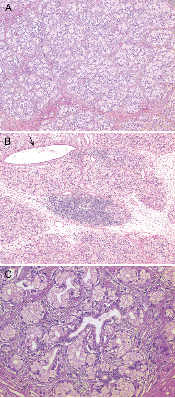
Figure 2.

Histological findings of the resected specimen. (A) Hyperplastic Brunner’s glands in lobules are separated by fibromuscular septa. HE stain, 40× (B) Lymphocytic infiltrate is observed and a lymphoid follicle is formed between lobules of Brunner’s glands at the center. Arrow indicates a cystically dilated duct. HE stain, 40× (C) Acini and ducts are well-preserved without nuclear atypia. HE stain, 200×.
