
Figure 4.
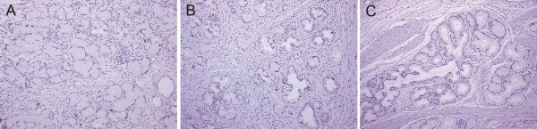
MIB-1 labeling in Brunner’s glands. Representative immunostain images of Brunner’s glands below surface epithelium associated with gastric foveolar metaplasia (A), below erosion (B), and below surface epithelium preserving intestinal nature (C) are shown. Brunner’s glands in (C) are rarely positive for MIB-1, compared to those in (A) and (B). All images are 100× magnification.