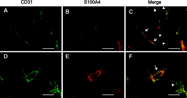
Fig. 1.
S100A4 expression in endothelial cells of intratumoral vessels. B16-BL6 cells were injected subcutaneously into C57BL/6 mice. Two frozen sections of the tumor tissue were double-immunostained with anti-CD31 antibody (a, d) and anti-S100A4 antibody (b, e). Merged imaged are also shown (c, f). Arrows and arrowheads in c and f indicate the examples of CD31+S100A4+ and CD31+S100A4− microvessels, respectively. Bars 200 μm

