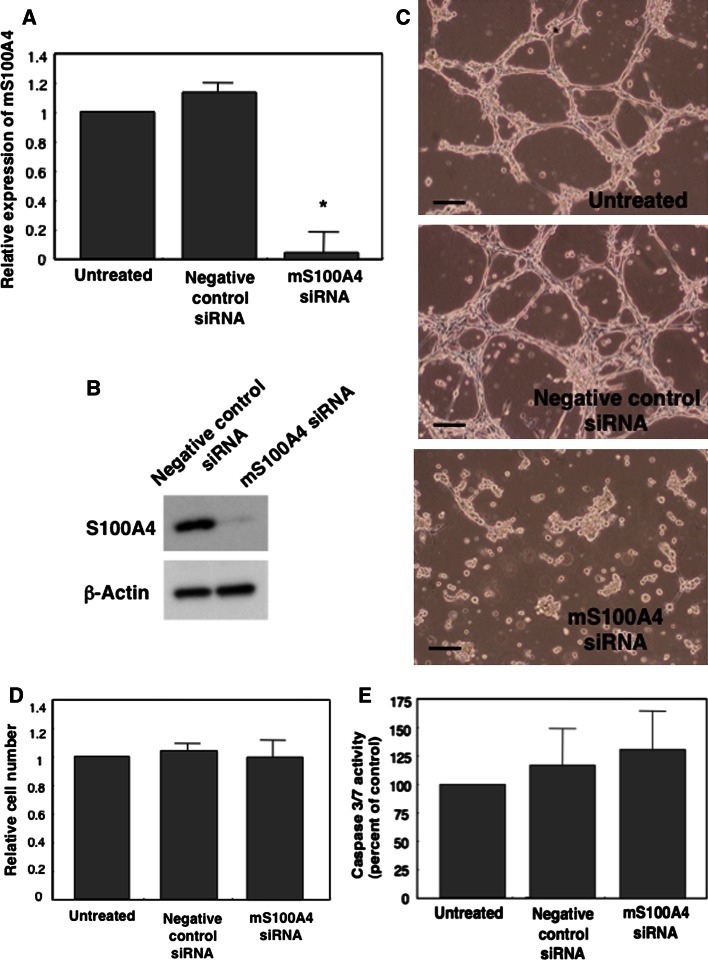
Fig. 2.
Anti-angiogenesis activity of S100A4 siRNA in vitro. MSS31 cells were transfected with murine S100A4 siRNA (mS100A4 siRNA) or control siRNA. Two days after transfection, 9.9 × 10⁴ MSS31 cells were seeded on ECM-gel thin-coated center-well dishes. RNA samples were extracted from transfected cells before seeding for real-time PCR analysis of S100A4 mRNA. HGF-induced capillary formation was assessed 16 h after Matrigel culture. a Relative expression of mS100A4 by real-time PCR analysis. *P = 0.01. b Western blot analysis of the expression of S100A4 in cells 24 h after siRNA transfection. Rabbit polyclonal anti-S100A4 antibody (Abcam, ab27957) and rabbit monoclonal anti-β actin antibody (Millipore, clone C4/MAB1501) were used. c Tube formation 16 h after siRNA treatment. Top, untreated control cells; middle, cells transfected with negative control siRNA; bottom, cells transfected with mS100A4 siRNA. Scale bars represent 50 μm. d Cell growth inhibition by mS100A4 siRNA. Cell growth was monitored by cell number analysis 16 h after siRNA transfection. e Caspase-3/7 activity. The activity was measured 16 h after siRNA transfection

