
Fig. 1.
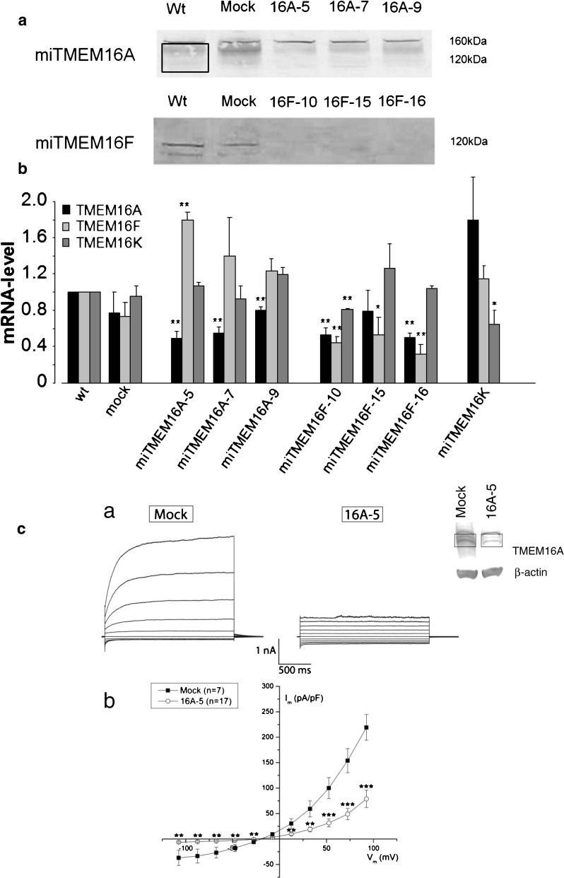
Verification of knock down of TMEM16A and F on protein and mRNA level. a SDS–polyacrylamide gel electrophoresis (10 % gel) of the three TMEM16A and the three TMEM16F knockdown clones compared to wild-type and mock clones. The membranes were stained with Ponceau Red (Sigma) as a control of identical protein loading in each lane. Membranes were incubated with primary antibody against TMEM16A; ab53212 (abcam) or TMEM16F; sc-136932 (Santa Cruz). TMEM16A is seen as a smear from 160–120 kDa (area inside the box). TMEM16A-5 and TMEM16A-7 show a clear downregulation of TMEM16A when compared to wt and mock, and in TMEM16A-9, a clear tendency towards downregulation is apparent. TMEM16F is seen as a band at 120 kDa and as a smaller isoform at 55 kDa. We were not able to quantify the protein bands because of the poor antibodies towards both TMEM16A and TMEM16F. The blot for TMEM16A is typical for N = 5, and the blot for TMEM16F is typical for N = 3. Five blots against TMEM16F were discarded because of too much unspecific binding. b Quantitative RT-PCR analysis of TMEM16A (black), F (light gray), and K (dark gray) relative mRNA levels in control, mock, three TMEM16A KD clones (TMEM16A-5,-7, and -9), three TMEM16F KD clones (TMEM16F-10, -15, and -16), and one TMEM16K KD clone. While a knockdown of TMEM16A led to an upregulation of TMEM16F, a knockdown of TMEM16F was accompanied by a downregulation of TMEM16A. Data are shown as means ± SEM, N ≥ 3, * P < 0.05, and ** P < 0.01. mRNA levels were normalized to the housekeeping genes acidic ribosomal protein and mouse β2 microtubulin. c Whole-cell membrane currents (Im) from ELA cells stably transfected with TMEM16A-miRNA and mock vector as indicated. a Each panel shows currents elicited at different membrane potentials in the range of −100 to +100 mV in steps of 20 mV in the presence of 1 μM free intracellular Ca2+. b Membrane currents measured at the end of voltage pulses (Vm) are plotted against the applied membrane potential after correction for the liquid-junction potential. The currents measured in miTMEM16A cells were significantly smaller than those of mock-transfected cells (**P < 0.01 and ***P < 0.001)