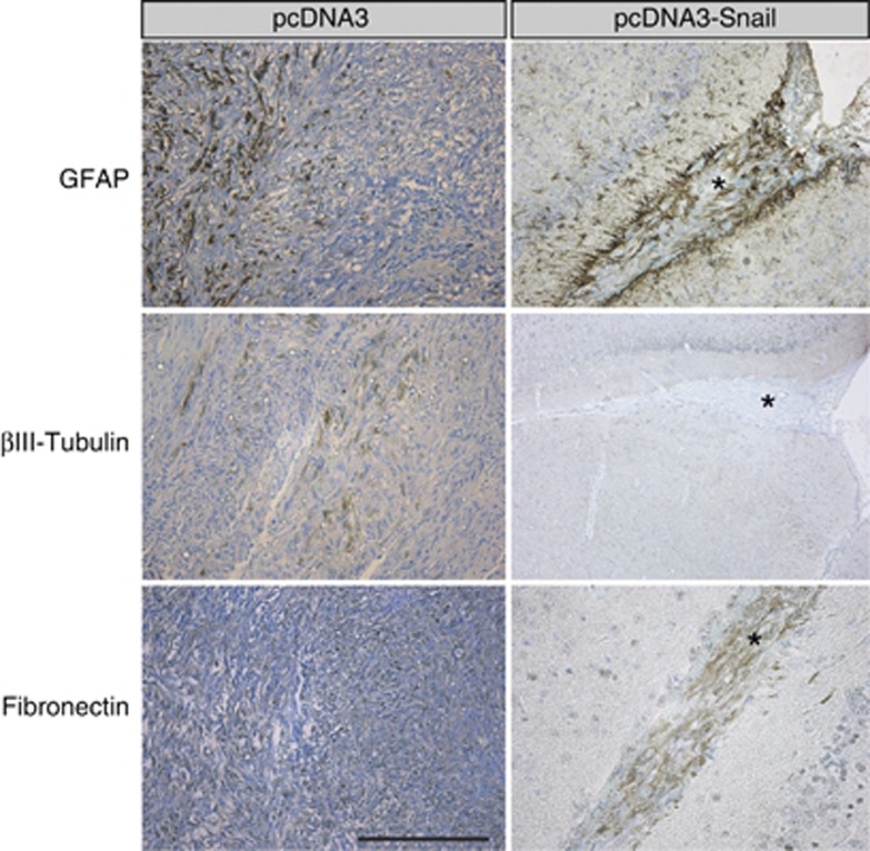
Figure 7.
Cell-specific marker expression in U-2987 MG invading tumor cells in vivo. Immunostaining for GFAP, βIII tubulin and fibronectin on tumor sections from mice injected with pcDNA3 or pcDNA3-Snail transfected U-2987 MG cells dissociated from gliomaspheres. Note the widespread tumor cell staining in the control tumors (left panels) versus the limited, streak-like formation of the invasive Snail-expressing tumors (right panels, stars). Scale bar: 200 μm.

