Figure 1.
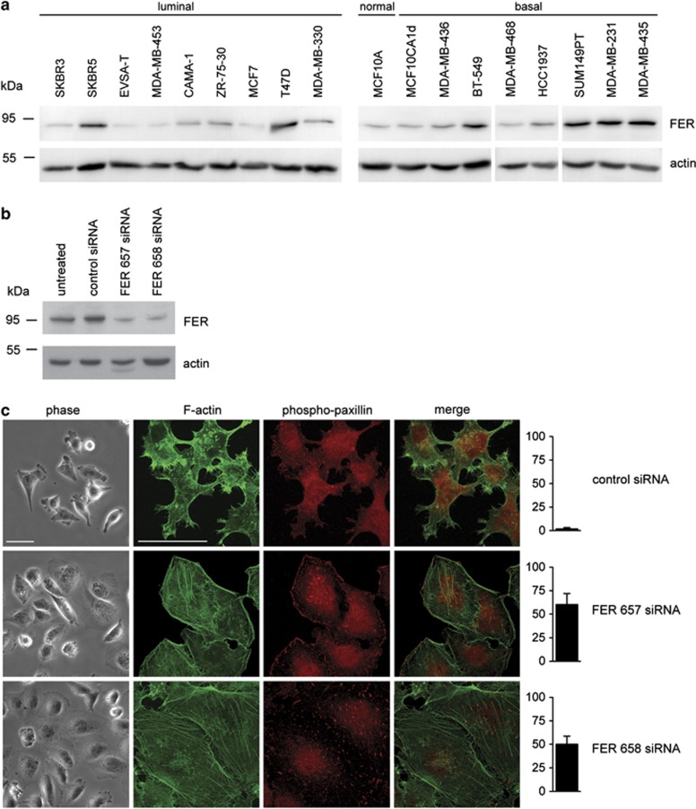
Inhibition of FER induces cell spreading, actin stress fibre and FA formation. (a) Total cell lysates of the indicated breast cancer cell lines were prepared and resolved by SDS-polyacrylamide gel electrophoresis. FER kinase expression was analysed by immunoblot. (b) KD of FER in MDA-MB-231 cells using siRNA transfection. FER expression was analysed by immunoblot 72 h after transfection. Actin was used as a loading control. (c) Control and FER KD MDA-MB-231 cells were plated on collagen I-coated glass coverslips. The morphology of live cells was analysed by phase contrast microscopy (phase). F-actin (green) and phospho-paxillin (red) distribution was analysed in fixed cells by immunofluorescence (IF) microscopy. The percentage of cells showing stress fibers is shown in the right bar graphs. Scale bar=50 μm. The results shown are representative of three independent experiments.

