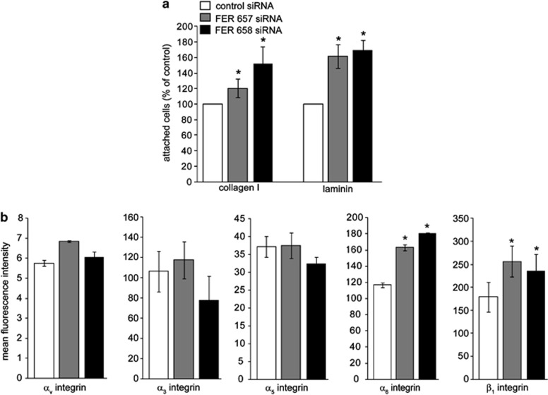
Figure 2.
FER regulates cell–matrix adhesion and integrin expression in breast cancer cells. (a) MDA-MB-231 FER KD cells were generated by transient transfection with the indicated siRNAs. FER KD cells were plated on 96-well plates coated with collagen I, fibronectin or laminin and the percentage of attached cells was determined. Values represent the relative proportion of attached cells ±s.e.m. * indicates significantly different proportions of adherent cells, relative to control (P<0.05, one-way analysis of variance (ANOVA)). (b) FER KD cells were trypsinized and surface expression of the indicated integrin subunits was analysed by fluorescence-activated cell sorting (FACS). Values represent mean fluorescence units ±s.e.m. * indicates significantly different integrin levels as compared with control (P<0.05, one-way ANOVA). The results shown are representative of three independent experiments.

