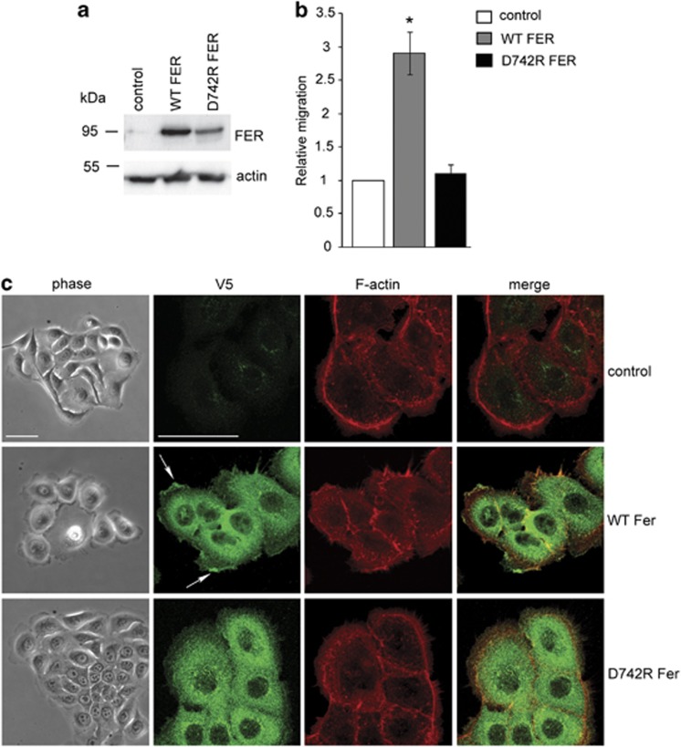
Figure 5.
FER regulates lamellipodia formation and migration. (a) The expression of V5-tagged wild-type (WT) and kinase-deficient (D742R) FER in stably transduced MCF10.CA1d cells was determined by immunoblot using an anti-V5 antibody. Actin was used as a loading control. (b) Control, WT and D742R FER-expressing MCF10.CA1d cells were seeded on collagen I-coated plates. The rate of migration was measured over 24 h using a modified wound-healing assay. Values represent the migration rate normalized to control ±s.e.m. * indicates significantly different migration rates relative to control (P<0.05, one-way analysis of variance (ANOVA)). (c) Control and WT or D742R FER-expressing MCF10A.CA1d cells were plated on collagen I-coated glass coverslips. The morphology of live cells was analysed by phase contrast microscopy (phase) after 24 h. Cells were then fixed and processed for immunofluorescence (IF) microscopy using an anti-V5 antibody to detect exogenously expressed FER-V5 (green) and Alexa 555-conjugated phalloidin to visualize F-actin (red). The arrow indicates accumulation of WT FER at the leading edges of lamellipodia. Scale bar=50 μm.

