Figure 6.
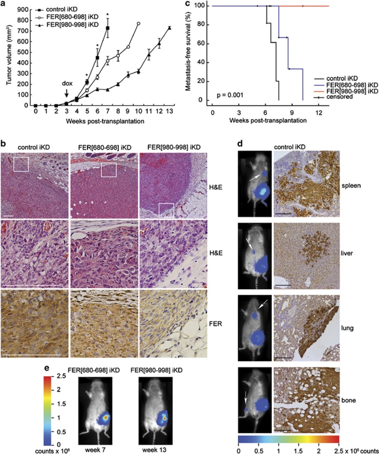
FER regulates breast tumour growth and metastasis. (a) Luciferase-expressing MDA-MB-231 iKD cells were orthotopically transplanted into recipient mice. Upon development of palpable tumours, mice were switched to a doxycycline-containing diet (dox; arrow) to induce shRNA expression. Values represent the mean tumour volume±s.e.m. *P<0.05, n=12 (control and FER (980–998) iKD), n=13 (FER (680–698) iKD). (b) Haematoxylin and eosin (H&E) and IHC staining for FER in primary tumours from mice transplanted with the indicated cell lines. Scale bar=200 μm. (c) Kaplan–Meier metastasis-free survival plot. Animals were monitored by bioluminescence and killed when distant metastases developed. (d) Representative images of mice from the control group at end point. Metastatic spread was assessed using bioluminescence imaging (left; arrow). IHC staining for vimentin was performed to confirm distant metastases of control tumours. Scale bar=100 μm. (e) Representative images of mice from the FER (680–698) and FER (980–998) iKD groups at end point.

