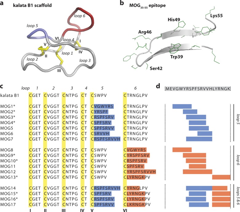
Figure 1.
Molecular grafting of antigenic peptides onto a cyclotide scaffold. (a) The cyclotide kalata B1 is stabilized by three conserved disulfide bonds (shown in yellow) and a head-to-tail cyclized backbone, which together form the cyclic cystine knot motif. The six conserved cysteines (numbered with roman numerals) divide the backbone into six loops, including loops 5 and 6 that are amenable to molecular grafting and colored in blue and red, respectively. (b) Native structure of the MOG35–55 bioactive epitope extracted from the three-dimensional structure of the entire MOG (myelin oligodendrocyte glycoprotein) protein. The epitope comprises two antiparallel β-sheets; selected residues are numbered in single letter code, and amino acid side chains are shown in green. (c) Aligned sequences of kalata B1 and novel grafted molecules MOG1–17. The six cysteine residues are highlighted in yellow, and the six loops are numbered. The grafted sequences in loops 5 and 6 are highlighted in blue and red, respectively. Grafted MOG peptides that adopted a native-like globular fold are marked with an asterisk. (d) The MOG35–55 bioactive epitope was grafted into the kalata B1 scaffold by dividing the full epitope sequence (shown on top) into smaller fragments and inserting them into either loop 5, loop 6 or loops 5 and 6.

