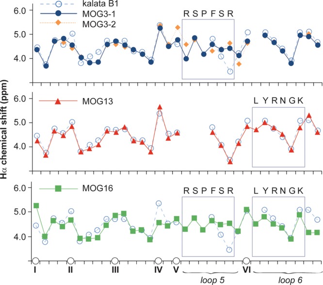
Figure 2.

Structural characteristics of MOG-grafted peptides. The Hα chemical shifts of MOG-grafted peptides and kalata B1 are shown. The positions of cysteines are indicated on the horizontal axis to align the sequence (non-cysteine residues are not shown for clarity). Two conformations for MOG3 (referred to as MOG3-1 and MOG3-2 in this figure) were observed. The different symbols used for each peptide are shown. Segments of the parent scaffold that have been grafted onto in loops 5 and 6 are boxed, and the sequences of the grafted epitopes are also shown.
