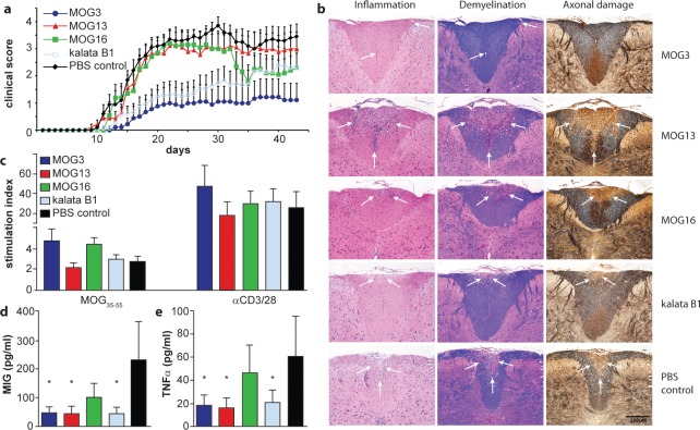
Figure 4.
Activity of novel grafted peptides in vivo in experimental autoimmune encephalomyelitis in mice. (a) Clinical score of EAE mice after vaccination with MOG-grafted peptides (MOG3, dark blue line; MOG13, red line; MOG16, green line) and controls (kalata B1, light blue line; PBS, black line) was monitored. (b) The influence of MOG-grafted cyclotide vaccination on the formation of CNS inflammatory and demyelinating lesions was examined by histological studies of fixed tissue using hematoxylin/eosin, Luxol fast blue, and Bielshowsky silver staining. Regions of inflammation, demyelination, and axonal damage are highlighted by white arrows. (c) Proliferation of spleen cells in response to the encephalitogen MOG35–55 and stimulation by the polyclonal activators, anti-CD3 and anti-CD28 antibodies. (d, e) Significantly reduced levels of the chemokine MIG (d) and TNFα (e) were demonstrated in non-stimulated spleen cell supernatants generated from animals treated with MOG3, MOG13, and kalata B1. * p < 0.05 compared to PBS control.

