
Figure 1. Expression of SSEA-4 in human SSCs.
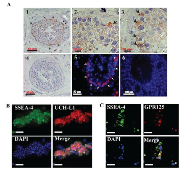
(A): Immunohistochemistry (A1-A4) and immunofluorescence (A5-A6) staining of SSEA-4 antibody on human testis paraffin sections. SSEA-4 is expressed by a limited number of cells (shown by arrows) located on the basement membrane of the seminiferous tubules where the SSCs are also located, a pattern that suggests that SSCs are a subpopulation of SSEA-4 (+) cells. (A4 & A6): Negative control stainings without primary antibody. Nuclei (blue) are stained with hematoxylin (A1-A4) or DAPI (A5-A6). Scale bars: 50 μm (A1, A4, A5, A6), 10μm (A2, A3), 100μm (A6) (B-C): SSEA-4 (+) cells were isolated from human testis using MACS and co-stained with anti-SSEA-4 and UCH-L1 (B), or GPR125 (C). The majority of cells co-expressed SSEA-4 and UCH-L1 or GPR125 also known as human SSCs markers, as shown in the merged images in (B) and (C). Nuclei were stained with DAPI (blue). Scale bars = 100μm.