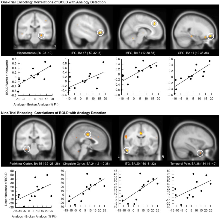
Figure 4.
Correlation of encoding-related brain activity with analogy detection at test. The location of the cluster of voxels, where correlations reached significance, is shown on anatomical brain images. BA, Brodmann area; IFG, inferior frontal gyrus; SFG, superior frontal gyrus; MFG, medial frontal gyrus; ITG, inferior temporal gyrus. Scatter plots of these correlations are presented below brain images. The first eigenvariate of the cluster of significantly correlating activity is displayed in arbitrary units on the y-axis. The difference in the percentage of fit responses to analogs versus broken analogs is displayed on the x-axis. All data come from good subliminal encoders (N = 12). The top panel shows correlations of activity increases to subliminal word pairs versus pairs of consonant strings during one-trial encoding with analogy detection. The bottom panel shows correlations of linear activity increases over the ninefold encoding of word pairs with analogy detection.

