
Figure 2.
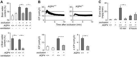
Reduced brain swelling, ICP elevation, and BBB disruption in AQP4−/− mice after BCAO. A) Brain water content was measured in nonventilated (as in Fig. 1A) and ventilated (as in Fig. 1D) CD1 mice at 10 and 15 min after 4.5 and 30 min of BCAO, respectively. Top panel: summary of brain water content for 5 mice/group. Bottom panel: difference in brain water content measured in BCAO vs. control mice. B) ICP was recorded between 0.5 and 10 min after 4.5 min of BCAO in nonventilated CD1 mice. Top panels: representative ICP recordings. Bottom left panel: mean ICP (average over 1–10 min after BCAO; n=4–5/group). Bottom right panel: difference in mean ICP (ΔICP) in BCAO vs. control mice. C) Evans blue extravasation measured in nonventilated CD1 mice at 10 min and 6 h after 4.5 min of occlusion (n=5/group). *P < 0.05, **P < 0.01.