
Figure 3.
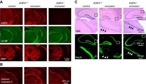
Reduced cellular damage in AQP4−/− mice after BCAO. A) AQP4, GFAP, and MBP immunofluorescence in brain sections of nonventilated C57bl/6 mice at 24 h after 30 min of BCAO. Representative of 4 mice/group. B) Immunofluorescence of cleaved caspase-3 (apoptosis marker) in control and in AQP4+/+ and AQP4−/− CD1 mice at 24 h after 5 min of BCAO (representative of 3 mice/group). C) H&E-stained hippocampal sections (top panels) in mice treated as in A, shown together with NeuN immunofluorescence (bottom panels) (representative of 3 mice/group). Higher-magnification images of boxed areas are shown under each image. Arrows indicate loss of NeuN immunofluorescence.