
Figure 5.
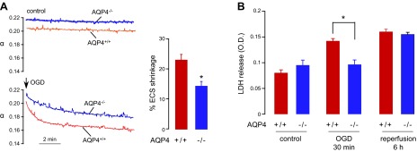
Reduced brain cell swelling and cell death in hippocampal slices from AQP4−/− mice after OGD. A) ECS volume was measured continuously in brain slices by a microfiberoptic fluorescence method. Left panel: representative data for hippocampal slices from AQP4+/+ and AQP4−/− mice without and during OGD. Right panel: percentage of ECS shrinkage, as calculated by relative ECS volume change [1 − (αOGD/αbasal)] (5 slices from 5 mice/group). *P < 0.05. B) LDH release from organotypic hippocampal slice cultures of AQP4+/+ and AQP4−/− mice after 30 min of OGD and at 6 h after return of slices to normal oxygen–glucose levels (reperfusion; 8 slices/plate insert, 4 inserts/group). *P < 0.05.