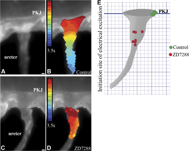
Figure 2.
HCN channel conductance is required for localizing the origin of UUT peristalsis to the PKJ. A–D) Videomicroscopy (A, C) and optical mapping (B, D) were used to analyze peristaltic and electrical activity in whole-mount urinary tract explants with and without HCN channel inhibition (see Supplemental Movies S1 and S2). Explants incubated with Tyrode's saline alone (A, B) or with the HCN channel inhibitor ZD7288 (30 μM; C, D) were loaded with the voltage-sensitive dye RH237. Isochronal maps (B, D) of membrane depolarization over time (red to blue) were recorded, with red demarking the initiation site of electrical excitation. Explants treated with Tyrode's solution alone exhibited unidirectional contractile (Supplemental Movie S1) and electrical waves that initiate at the PKJ (red) and propagate distally down the renal pelvis and ureter (yellow to blue) (A, still image; B, isochronal map). In contrast, explants treated with ZD7288 exhibited twitch-like contractions (Supplemental Movie S2) and desynchronized electrical activity that initiated in random segments of the UUT distal to the PKJ (C, still image; D, isochronal map). E) Quantification of data analyzing the effect of HCN inhibition on the origin of UUT contraction. Origin of contraction in control explants localized to the PKJ (green circles) in 7 of 7 explants. In contrast, the origin of contraction in all 7 explants incubated with ZD7288 was shifted to random sites of the UUT, distal to the PKJ (red circles). Scale bars = 100 μm.

