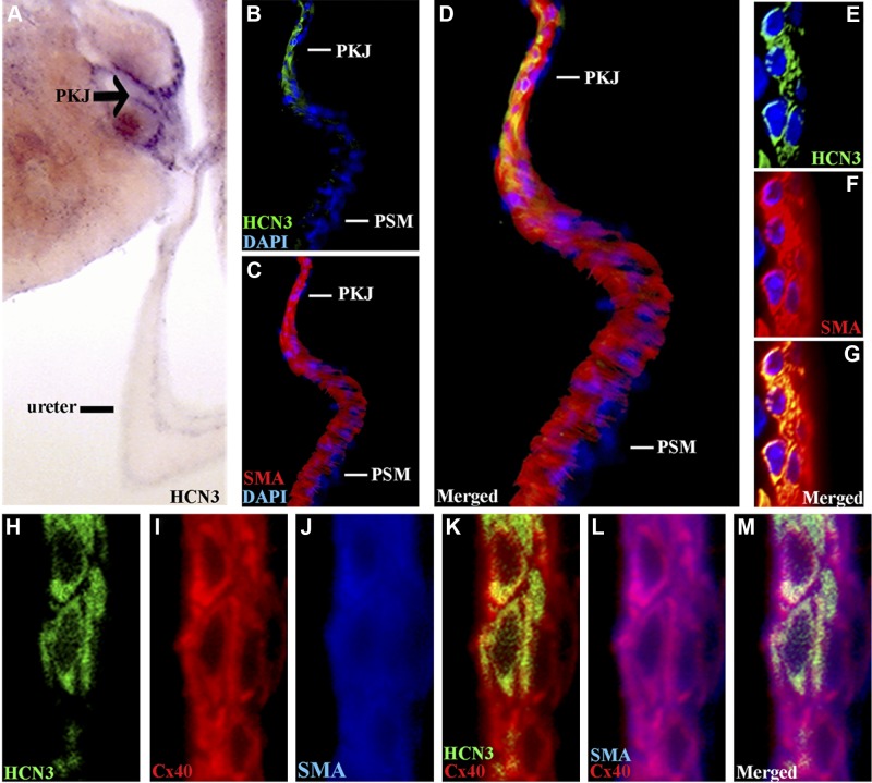
Figure 5.
HCN3-expressing cells are localized to the PKJ and coupled to adjacent smooth muscle by gap junctions. A) Chromagenic immunohistochemistry on 80-μm UUT sections demonstrates that HCN3 (purple reaction product) is highly expressed and localized to the PKJ (arrow). B–D) Triple immunofluorescent staining for SMA (red; C, D), HCN3 (green; B, D), and DAPI (blue) demonstrates that HCN3-expressing cells (B) are integrated within the UUT smooth muscle coat at the PKJ (C), but not the thicker pelvic smooth muscle (PSM) of the more distal UUT (D, merged). E–G) Confocal microscopy of the PKJ demonstrates that HCN3+ cells (green; E) are adjacent to and in direct contact with the smooth muscle (red; F, G; G, merged). H–M) Triple immunofluorescent staining for HCN3 (green; H, K, M), Cx40 gap junction (red; I, K–M) and SMA (blue; J, L, M) demonstrates that HCN3 (H) cells of the PKJ are coupled to adjacent smooth muscle (J) by Cx40 (I), which is coexpressed by both cell populations (K, HCN3/Cx40 merged; L, SMA/Cx40 merged; M, all merged).

