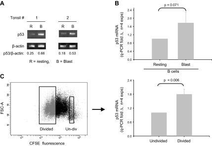
Figure 2.

B cells replicating in response to BCR:CD21-L, IL-4, and BAFF exhibit elevated levels of p53 mRNA. A) Semiquantitative RT-PCR analysis of p53 mRNA levels in resting B cells and activated d 4 cultures stimulated with BCR:CD21-L, IL-4, and BAFF. Values represent ratios of densitometric intensity in the p53 and β-actin bands B) qPCR assessment of p53 mRNA in resting B cells vs. d 4 blasts. ΔCt values for p53 were obtained by comparisons with β-actin. Values for fold difference (Δ) were obtained by comparing ΔCt values in activated cultures with the ΔCt values of B cells prior to activation. P value shows that the differences between Δ values in resting vs. activated cells in a total of 4 replicate experiments were of borderline significance, using a 2-tailed paired Student's t test. C) Undivided blasts and divided blasts within activated cultures were sorted on the basis of CFSE fluorescence. Left panel: representative experiment. Right panel: results for p53-specific q-RT-PCR of isolated RNA. Divided blasts express significantly more p53 mRNA than do undivided blasts within the same cultures (P=0.006; 2-tailed, paired Student's t test).
