
Figure 3.
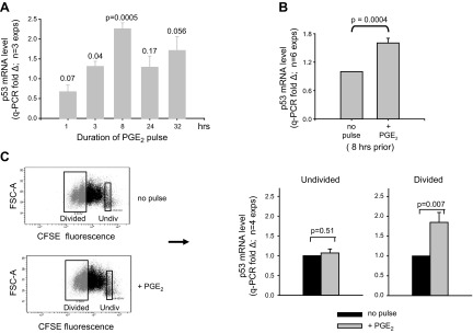
PGE2 augments p53 mRNA expression during TI clonal expansion. A) B-cell cultures activated with BCR:CD21-L, IL-4, and BAFF were pulsed with exogenous PGE2 (50 nM) or ethanol vehicle alone on d 4; mRNA was isolated at varying periods after the pulse. qPCR of cDNA was used to measure the levels of p53 mRNA in PGE2-pulsed and control cultures. Values for fold difference (Δ) were obtained by comparing ΔCt values in PGE2-pulsed cultures with the respective ΔCt values in vehicle-pulsed cultures at the same time point. Dotted line represents a value of Δ = 1 for each of the vehicle control cultures. Results are the mean ± se Δ fold difference in 3 separate experiments, where P represents significance of differences between Δ values in PGE2-pulsed vs. vehicle-pulsed cultures. B) Pooled results from a total of 6 experiments which evaluated p53 mRNA levels in cultures activated as in A with/without an 8 h pulse of PGE2 on d 4. C) Day 4 activated cultures of CFSE-labeled cells with or without pulse of exogenous PGE2 8 h earlier were harvested and sorted on the basis of their division status: undivided vs. divided (3–4 divisions). qPCR of cDNA with p53 and β-actin probes was used to monitor p53 mRNA levels, as in Fig. 2. Values for Δ in each sorted population were obtained by comparing ΔCt values in cells from PGE2-pulsed vs. unpulsed cultures. P values show that a pulse with PGE2 significantly augments p53 mRNA levels in the divided blasts (P=0.007) without affecting p53 levels in the undivided cells (P=0.51; 2-tailed, paired Student's t test).