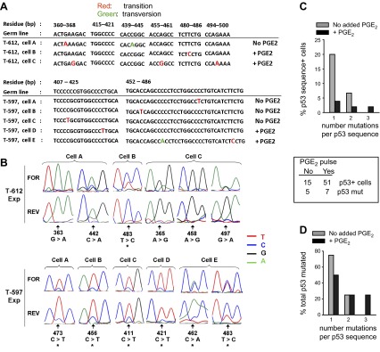
Figure 9.

p53 mutations are present within dividing lymphoblasts of a TI response to BCR:CD21-L, IL-4, and BAFF with or withouth supplementary PGE2. A) p53 amplicons from experiments in Fig. 7 were sequenced (see Materials and Methods). Shown are sequenced segments with identified mutations from 8 distinct cells from 2 donors, as well as the corresponding germline p53 sequence (numerical positions represent residues of germline DNA beginning at the transcriptional start site). Transition mutations are in red; transversion mutations are in green. B) Segments of forward (FOR) and reverse (REV) p53 sequence chromatograms indicating the mutations detected per individual cell. Arrows indicate mutated residues. Asterisks indicate cells in which both the mutation (predominantly expressed) and the original wild-type sequence (minor) were coexpressed. Coexpression was validated by similar results on sequencing in either direction. C) Summarized data showing the percentage of total p53 sequence-positive single cells that expressed 1, 2, or 3 mutations/sequence. D) Normalized data from panel C, showing the percentage of total p53-mutated cells that expressed 1, 2, or 3 mutations/sequence. For both C and D, statistical power is too low to determine significance.
