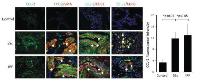
Figure 4.

Left, Immunofluorescence staining with anti-CCL2 antibodies in lung tissue from normal controls and patients with SSc or IPF. Arrows indicate double-positive cells. Original magnification × 400. Right, Fluorescence intensity of CCL2 in the 3 groups. Bars show the mean ± SD. See Figure 3 for definitions.
