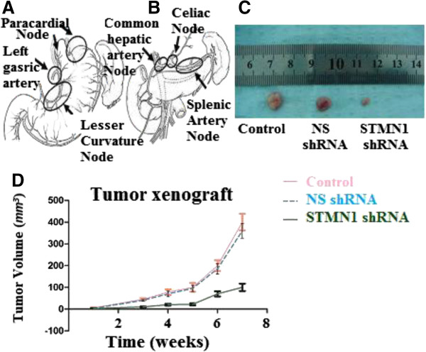
Figure 6.

Abdominal lymph node clearance and xenograft tumor models in nude mice. (A) Perigastric lymph nodes, lymph nodes along the left gastric artery and lesser curvature lymph nodes dissection. (B) Distant abdominal lymph node dissection. Abdominal Esophagus drains into superior gastric artery, celiac axis, common hepatic artery and splenic artery lymph nodes. (C) Esophageal cancer cells were either untreated or transfected with Non-silencing shRNA (scrambled sequence) as a negative control and transfected with STMN1 shRNA were xenografted subcutaneously in the BALB/c-nu/nu male mice. Tumor mass (xenograft) volume was measured every week from week 3 to week 7. Data are expressed as percentage change (Means ± S.D.) compared with controls and represent four independent experiments. (P < 0.05 vs Non-silencing shRNA, one-way analysis of variance (ANOVA) followed by Tukey’s multiple comparion). (D) Photograph of xenografts dissected from nude mice after 7 weeks subcutaneous inoculation showing suppression growth of cancer cells transfected with stathmin1 shRNA as compared to cells transfected with untreated or transfected with Non-silencing shRNA.
