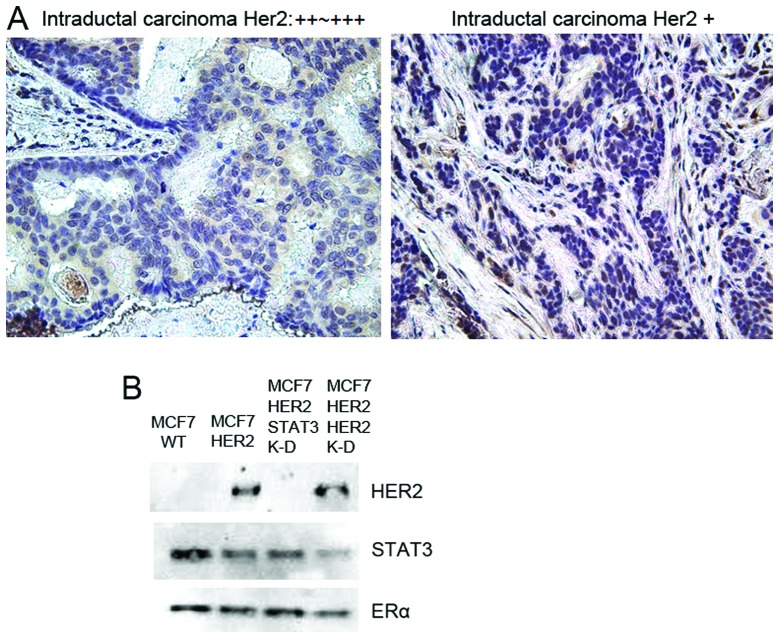
Figure 6.
STAT3 activation in HER2-overexpressing, ER-positive human breast cancer. (A) Tissue microarray of human breast cancer patients. Breast cancer patient tissue microarray slides (BRC961) were obtained and stained using standard immunohistochemistry method with an IHC validated phospho-STAT3 antibody (Y705). pSTAT3-positive patient tissues are shown. Intraductal carcinoma HER2++-HER2+++, invasive ductal carcinoma HER2+. (B) Immunoprecipitation revealed the interactions between HER2/ER and STAT3. To examine the physical binding of ER, immunoprecipitation was performed with ERα antibody in MCF7-HER2 cells. For MCF7-HER2 cells, STAT3 K-D is STAT3 knockdown and HER2 K-D is HER2 knockdown cells. After IP, pellets were resolved on a PAGE gel and immunoblotted for HER2, STAT3 and ER proteins.

