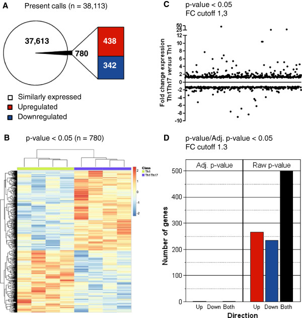
Figure 2.
Identification of differentially expressed genes in Th1Th17 vs. Th1 cells. Matched Th1Th17 and Th1 subsets from four HIV-uninfected donors were sorted and stimulated as in Figure 1. Total RNA was extracted and reverse transcribed into cDNA that was then hybridized on the Human Genome U133 Plus 2.0 Array (Affymetrix). Statistical analysis using one-way ANOVA was performed to identify differentially expressed genes (p-value <0.05) and the differential expression fold change (FC) was calculated. (A) Shown is a schematic representation of the number of ”present calls” shared (n = 38,113) and differentially expressed (n = 438 upregulated and n = 342 downregulated) in Th1Th17 vs. Th1 (p-value <0.05). (B) Hierarchical clustering analysis of differentially expressed probe sets separated the 8 samples in two groups corresponding to Th1Th17 and Th1 subsets. (C) Depicted is the fold change of differentially expressed genes in Th1Th17 vs. Th1 (p-value <0.05 and FC cut off 1.3). (D) Shown are numbers of probe sets differentially expressed in Th1Th17 vs. Th1 with a fold change cut-off of 1.3 according to their p-value (n = 265 upregulated in red and n = 235 downregulated in blue) or adjusted p-value (n = 2 upregulated).

