
Fig. 1.
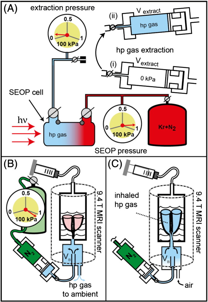
Hp krypton extraction and transfer from the SEOP cell, operating at 90–100 kPa, to the lungs at ambient pressure. (A) A pre-evacuated volume Vextract = 790 cm3 in the extraction unit (i) was filled to approximately 6 kPa during hp gas extraction (ii). (B) The extraction unit was moved to the MRI scanner and the N2 gas operated piston pressurizes the hp gas mixtures to a pressure slightly above ambient. The hp gas was then pushed through connecting tubing into a storage container (VB). The lung was located upside down in glucose solution within the breathing apparatus with the trachea connected to VB. (C) A slight suction on the breathing apparatus (substituting for the pleural cavity) caused the lung to expand and to inhale the hp gas.