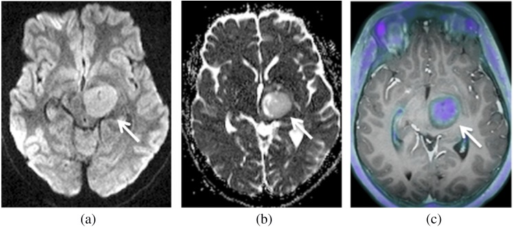
Figure 2.

Images of an 11-year-old female with pathologically proven low-grade glioma (arrows). Axial image: (a) diffusion weighted image, (b) apparent diffusion coefficient (ADC) map and (c)18F-choline-PET/T1 MR fused. Moderate 18F-choline uptake and mildly reduced ADC is present within the tumour.
