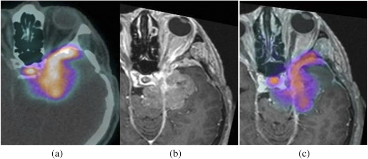
Figure 3.
Images of a 47-year-old male with a skull base meningioma following surgery, being considered as a candidate for radiotherapy. Positron emission tomography (PET)-CT images (a) demonstrate a 68Ga-DOTATATE (derivative of octreotide)-avid lesion in the left middle cranial fossa. T1 post-contrast MR images (b) show anatomical detail of the tumour location. Combined PET-MRI (c) allows visualization of the full extent of this disease.

