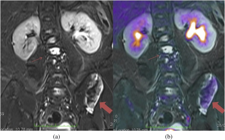
Figure 9.
(a) Coronal short tau inversion–recovery MRI shows multiple areas of increased abnormal signal in the vertebra (thin arrows) and left iliac wing (thick arrows). (b) Active disease is differentiated from stable disease on the combined positron emission tomography-MRI as areas of increased 18F-fludeoxyglucose uptake.

