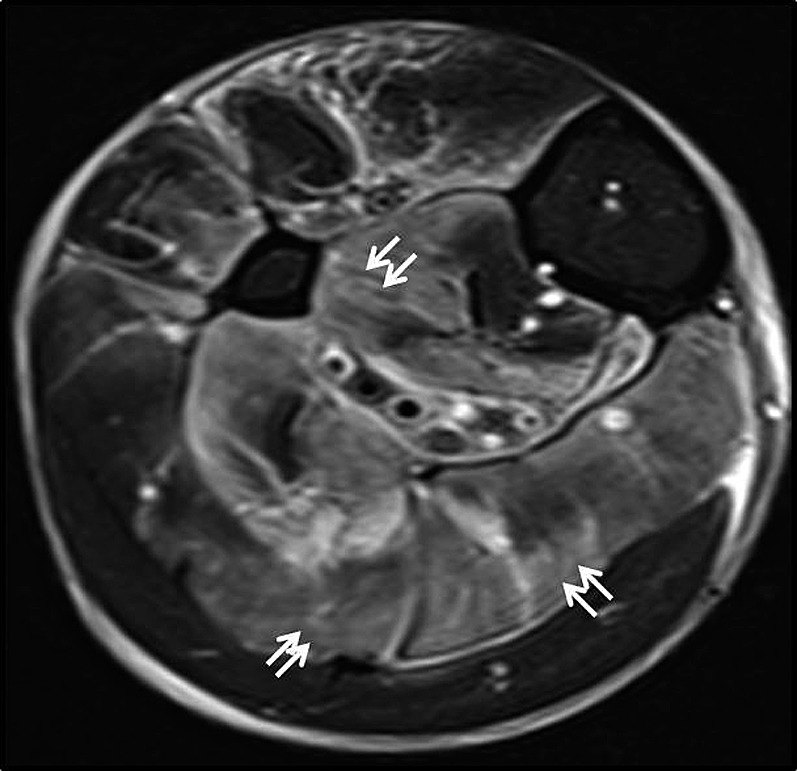
Figure 10.

Infective myositis. Axial fat-suppressed T2 weighted MR image shows patchy areas of T2-hyperintense signal involving the soleus muscle (superficial posterior compartment) and also the deep posterior muscle compartment (white arrows). No thick (>3 mm) T2-hyperintense signal was evident in the deep intermuscular fascia. Findings were compatible with myositis. Patient was managed conservatively with antibiotics.
